Introduction and Research Perspective

Posted by Tommy


The goal of our research program is to discover how system-level properties of large groups of eukaryotic and prokaryotic cells emerge from microscopic dynamics, both at single-cell and molecular scales. Single cell behavior is well understood in eukaryotes and prokaryotes, but it is their behavior in large communities that is important to multi-cellular life. For all eukaryotic and prokaryotic multi-cellular organisms, life depends on the coherent functioning of large cell aggregates. In traditional condensed matter physics, great progress is made by discovering how bulk system properties arise from the behavior of the sub-units. If the same is true of living condensed-matter, then an understanding of the relationship between the cellular sub-units and the function of their collections is crucial to understanding life in multi-cellular organisms and, as a consequence, controlling it.

Large cell groups that function collectively, such as tissues and bacterial biofilms, often have sub-groups with specialized jobs. This heterogeneity in cell function is most dramatically illustrated by comparing cells at the surfaces to cells within the bulk. For example, in stratified epithelia, the apical and basal layers of cells have totally different functions than the inner lateral cells. Likewise, in bacterial biofilms, cells at the interface between the colony and nutrients are always the most metabolically active, and cells far away from nutrient sources must rely on the metabolic intermediates of surface cell groups. Thus, to understand collective behavior of multi-cellular systems as wholes, our research program focuses on eukaryotic and bacterial cell layers on surfaces, as well as many types of bulk three-dimensional cell aggregates.

It is impossible to see deep into thick 3D cell aggregates with visible light due to strong multiple scattering and absorption. To surmount these imaging problems, we are building new tools that use radiation at wavelengths that pass through biological materials with, primarily, single-scattering events: x-rays and IR illumination. These tools are currently being developed and, thus, are top-secret. If you would like to see them in action, come pay us a visit!

We also study cells on surfaces, which can be imaged with visible light, allowing the use of ordinary fluorescence microscopy. Throughout our research program, we compliment micro-scale studies macro-scale measurements involving low-resolution time-lapse imaging and bulk-rheological studies. At the molecular-scale, we do x-ray scattering to explore bio-molecular self-assembly to understand the interactions between the many extracellular materials, as well as their interaction with cell surfaces.
Soft Matter Manufacturing

Updated August 24, 2017Posted by Chris O'Bryan


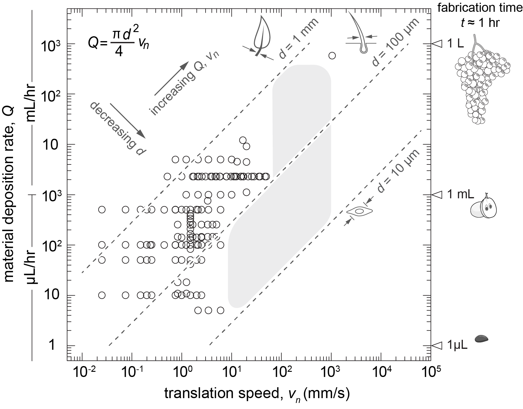
Manufacturing complex structures such as living tissues and organs requires large structures to be printed with high feature resolution over short time periods. To achieve this goal, a balance must be obtained between printer speed, manufacturing time, print volume, and the feature size of the printed structure. We can approximate this balance between speed, time, volume and resolution through the relationship Q = vnπd2 / 4 where Q is the material deposition rate, d is the feature diameter, and vn is the nozzle translational speed. Thus, a given feature size can be described by a combination of material deposition rates and nozzle translational speeds, creating a single manufacturing curve in Q-vn space. In our recent MRS review article, we plot a family of manufacturing curves corresponding to feature diameters between 10 µm and 1000 µm in Q-vn space along with data collected from a literary survey of 3D printing methods with sacrificial materials demonstrating the achievable manufacturing space. Although significant progress has been made in the field of tissue engineering, further advancements in 3D printing are necessary before large structure can be achieved with high resolution over short time periods.

Soft Matter 3D Printing

Updated March 28, 2017Posted by Chris O'Bryan


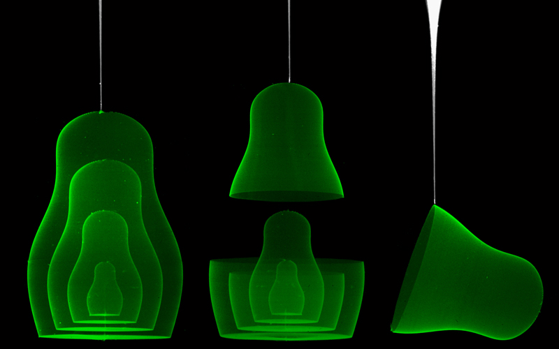
A major challenge in soft matter engineering is the precise manufacturing of soft delicate materials into complex constructs. Here at the University of Florida, we leverage the unique rheological properties of jammed granular microgels, specifically their low yield stress, short thixotropic time, and spontaneous reflow after yielding, to 3D print soft matter structures. Although these jammed microgel systems contain >95% solvent, they behave as a solid at low stress, exhibiting a rate-independent yield stress and a relatively flat elastic shear modulus that dominates over a wide range of frequencies. Thus, we often refer to these jammed microgels as liquid-like solids. Our lab has demonstrated the ability to 3D print hydrogel, silicone, and cellular structures using these liquid-like solids as support and continues to develop new jammed microgel systems to improve printing performance by reducing interfacial effects and reducing charge density.

Intercellular force transmission by contraction-generated pressure driven flow

updated January 9, 2012Posted by Steven Zehnder


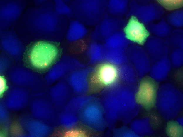
Cell force generation and transmission are essential to cell-to-cell interactions and cell locomotion. Contraction of the cell's cytoskeletal network generates forces that can be transmitted directly to other cells by cell-cell adherens junctions or through a substrate by traction forces. Within monolayers, cytosolic fluid is transferred between cells through gap junctions. The coupling between intercellular fluid movement and cytoskeletal contraction can give rise to a different type of cell-cell force transmission, in which cells pump fluid into their neighbors. To study intercellular hydrostatic force transmission, we monitor the movement of fluid between cells and we measure the associated changes in cell volume. MDCK cells are labeled with fluorescent cytosol dyes, and we monitor the cells with time-lapse microscopy.

MDCK cells are dyed and imaged in time-lapse fluorescence microscopy to monitor intercellular fluid transfer (green) and cell volume (blue).

3D collective cell mechanics

updated January 9, 2012Posted by Jolie Breaux


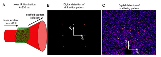
Mechanical cell behavior is vital to tissue health and dynamic cellular processes such as wound healing, and angiogenesis. There are many methods for measuring single cell and multi-cell mechanics on 2D surfaces. For example, traction force microscopy (TFM) is often used to measure cell generated forces while mechanical testing methods such as atomic force microscopy (AFM) are employed to determine materials properties of cells. These and other extant cell mechanics methods are optimal for single, isolated cells cultured on flat, 2D surfaces. These methods have been modified and applied to confluent cell monolayers, which are better models for many tissues than single cells. However, most cells in tissues are parts of 3D structures, not monolayers, so the development of new cell mechanics techniques in 3D systems is essential to elucidate cell mechanics in tissues. To study collective cell behavior in large, 3D systems, we have built a dynamic small angle light scattering (DSALS) system with environmental control for live-cell experiments. To penetrate deep into model tissue aggregates without multiple scattering events or significant attenuation, we illuminate our samples with near-infrared (NIR) light. The multi-cellular spatial distribution and dynamics within 3D scaffolds are determined through digital image analysis of scattering patterns.

(A) SALS is used to determine the distribution and dynamics of cells within bioscaffolds. (B) Periodic materials yield Bragg peaks, and amorphous samples (C) produce isotropic scattering.