Image Library
(Under Construction)
Scanning electron micrographs (SEMs)
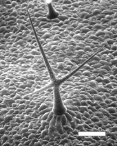 |
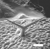
Mature wild-type trichomeSEM of a mature wild-type trichome on an Arabidopsis leaf. Scale bar is 100 µm. |
 |
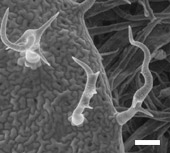
itb1 leafEnvironmental scanning electron microscopes can visualize living hydrated samples including live Arabidopsis plants. This ESEM shows developing trichomes on an itb1 mutant leaf. Scale bar is 100 µm. |
 |
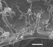
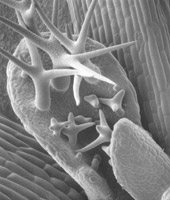
itb1 trichomesESEM of mature trichomes on an itb1 mutant leaf. ITB1 encodes a regulator of actin polymerization. Mutations in ITB1 cause cell expansion defects in trichomes and epidermal pavement cells. Scale bar is 50 µm. |
 |
Developing itb1 trichomeESEM of a developing trichome on an itb1 mutant leaf. Scale bar is 20 µm. |
 |
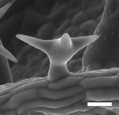
Mature frc2 trichomecryo SEM of a mature trichome on a frc2 mutant leaf. FRC2 encodes a katanin protein that regulates microtubule organization and cell expansion. Scale bar is 115 µm. |
Tissue Sections
 |
Nuphar flower bud 5x |
 |
Nuphar flower bud 10x |
 |
Nuphar flower bud 20x |
 |
Nuphar flower bud 20x |
Confocal Images
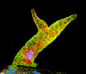 |
itb1 trichomeComposite confocal image of a developing trichome on an itb1 mutant plant. The trichome was treated with fluorescently labeled antibodies (green for microtubules and red for actin) and a dye that stains the nucleus blue. |