Thalassemia
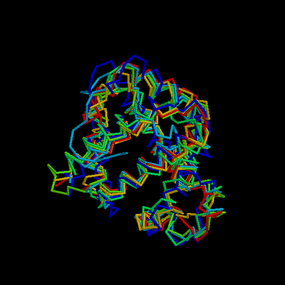
Thalassemia, also known as Cooley’s anemia, or Mediterranean anemia, is an inherited type of hemolytic anemia. caused by a variety of genetic defects in the gene coding for the structure of the globin chains of hemoglobin (Ortner, 2003). This type of disorder consists of the limited ability of the body to synthesize the beta chain of the globin molecule. This type of anemia is mainly found within central and eastern Mediterranean populations. Three manifestations of the disease occur; thalassemia major, in which bone changes always develop, and can be seen in radiographs, thalassemia intermediate may show them, but thalassemia minor does not (Ortner, 2003). The most severe bone changes occur in the skull, where the diploe of the cranial vault expands, leading to unusual thickness (Aufderheide, 1998).
Unlike iron deficiency anemia, thalassemia can produce post-cranial bone changes, sometimes identifiable in paleopathological contexts. The diameter of ribs is expanded, vertebrae show decreased height, increased width, cupping of end plates, and porosity. Finally, multiple line of arrested growth are present, due to multiple instances of inhibited growth, and delayed epiphysial closure seen in this disease. Thalassemia does not create any type of circulatory obstruction, nor does it have increased chance of osteomyelitis (Maggio, 2002).
Although this disease confers a degree of protection from malaria for carriers of the gene, it has a high degree of mortality for the sufferers. To survive, many people with thalassaemia need blood transfusions at regular intervals. Repeated transfusions cause the accumulation of iron in the heart and liver. If this condition is left untreated, it invariably leads to death by heart failure(Maggio, 2002).
