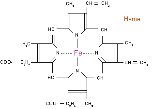
Anemia is referred to a lack of red blood cells– erythrocytes, which is the oxygen-carrying cell in the blood that contains the pigment hemoglobin. Hemoglobin is the iron-containing protein found in erythrocytes produced in the bone marrow, that combines with oxygen from the lungs and is carried and distributed to the cells and tissues of the body. There are several kinds of anemia, produced by a variety of underlying causes. These types are classified by the size of the red blood cell present in the body: decreased size(microcytic), normal size(normocytic) or enlarged size (macrocytic or megaloblastic).
Microcytic Anemia
The most common type of microcytic anemia (where blood cells measure under
under 80 femtoliters) is iron-deficiency
anemia. Other types of microcytic anemias result from hemoglobinopathies,
which are genetic defects that result in the abnormal structure of one
of the globin chains of the hemoglobin molecule. The most common of these
disorders are sickle cell anemia and thalassemia.
 Normocytic
Anemia
Normocytic
Anemia
Normocytic anemia (blood cell size of 80-100 fl), is primarily caused
as a side effect of acute blood loss, renal failure, or liver failure.
A rarer type of normolytic anemia is aplastic anemia, or bone marrow failure,
caused by the inability of the bone marrow to produce blood cells. Aplastic
anemias are much rarer than dietary deficiency or genetic defect anemias,
and progress much more rapidly.
Macrocytic Anemia
Macrocytic anemia (blood cell size greater than 100 fl) is most commonly
caused by a deficiency on vitamin B12, folic acid, or both. This is due
to inadequate intake or absorption. Pernicious anemia is caused by is
an autoimmune condition where the body lacks intrinsic factor, a glycoprotein
produced by cells in the stomach, which is required to absorb vitamin
B12 from food. Another way to obtain macrocytic anemia is chronic alcoholism.
Paleopathology of the Anemias
The paleopathology of the first agricultural
populations has created alot of interest recently, since the answer to
the question of whether or not the advantages of an agricultural way or
life outweight the disadvantages can be easily found in the remains of
those populations. Most skeletal changes involved with anemia consists
of cranial vault bone thickening, trabecular bone coarsening, thinning
of the cortical bone, and metaphyseal widening (Ortner, 2003). J. L. Angel
hypothesized that the diploic thickness present in crania diagnosed with
anemia must reflect a red cell production by the hyperostotic bone almost
twice of the normal diploe (J. L. Angel, 1966)